


For the XRD test of bulk samples, the bulk sample is often ground into an isotropic powder state, and then the sample is tested and analyzed by diverging linear X-rays. However, it is not practical for some bulk samples to be ground into powder, and micro-XRD analysis technology provides a powerful tool for such samples to analyze the original state of the sample and carry out phase analysis of various parts of the sample. In this paper, a geological sample lapis Lazuli is taken as an example to further illustrate the effect of micro-diffraction.

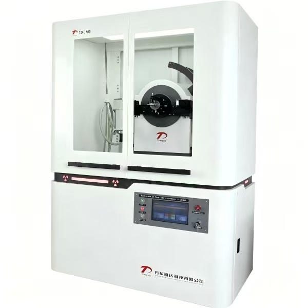
FIG1

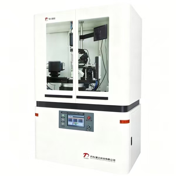
FIG2
![]()

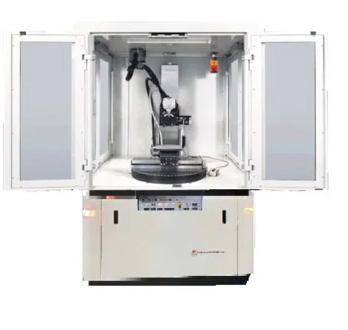
FIG3
2D microdiffraction is used to better characterize the structure of lapis lazuli. In situ measurements of lapis lazuli crystals and quartzite matrix were made using 0.5 mm spot spot and EIGER2 detector 2D mode (FIG. 1, FIG. 3). During the measurement, the sample rotates at phi of 3rpm and oscillates at psi from 0° to 35° for more reflection. Microdiffraction confirms that the matrix is predominantly fine-grained quartz (blue circles), while lapis lazulis crystals (red circles) contain symbionts of polycrystalline diaspore, metalumite, and gypsum (Figure 4).

FIG4