

- Home
- >
News
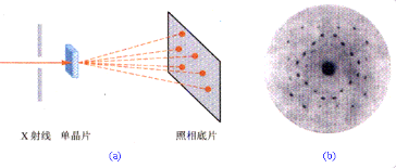
In 1912, Laue et al. predicted by theory and confirmed by experiment that diffraction can occur when X-ray meets crystal, proving that X-ray has the property of electromagnetic wave, which became the first milestone in X-ray diffraction.
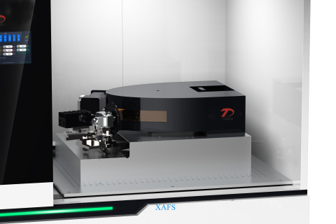
XAFS, as an advanced characterization technique for the local structure analysis of materials, can provide more accurate atomic structure coordination information in the short-range structure range than X-ray crystal diffraction.

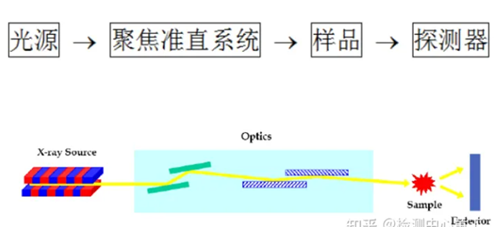
X-ray diffraction is a commonly used non-destructive analytical technique that can be used to reveal the crystal structure, chemical composition, and physical properties of substances.
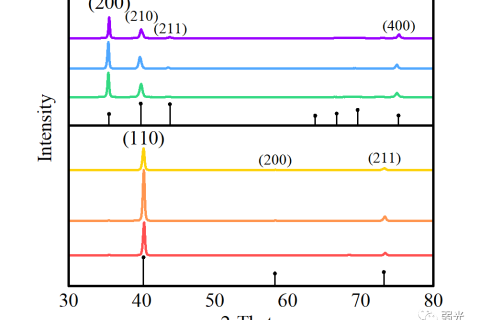
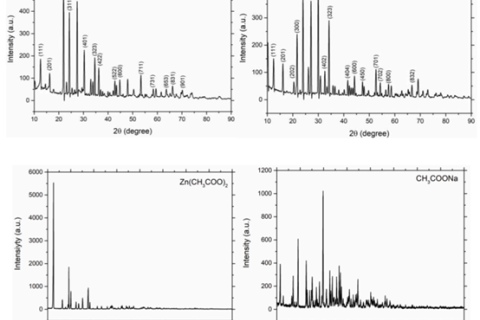
X-ray diffraction, through the X-ray diffraction of a material, the analysis of its diffraction pattern, to obtain the composition of the material, the structure or shape of the atoms or molecules inside the material and other research means.
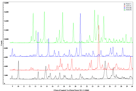
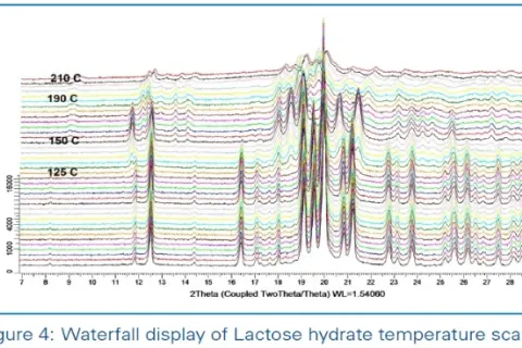
X-ray diffraction runs through every stage of drug quality control, such as the study of raw materials and preparations.
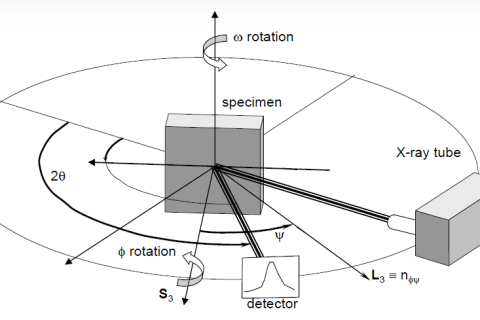
Residual stress has a great impact on the dimensional stability, stress corrosion resistance, fatigue strength, phase change and other properties of materials and components. Its measurement has been widely concerned by academia and industry.
XRD, is short for X-ray diffraction, as a material person, no matter what material is done, XRD is the most commonly used, the most basic means of characterization.
When a beam of extremely thin X-rays passes through a material with an uneven electron density of nanometer size, the X-rays will spread out in a small angular region near the direction of the original beam, this phenomenon is called small-angle X-ray scattering.
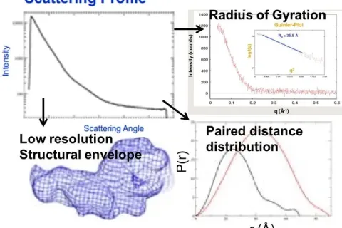
Small Angle X-ray diffraction (SAXD) is mainly used to determine the spacing of very large crystal faces or the structure of thin films.
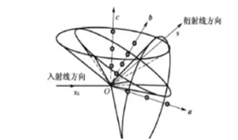
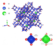
The use of X-rays to study the structure of crystals mainly through the X-ray diffraction phenomenon in the crystal.
X-ray diffraction technique is an analytical method used to study the structure of a substance. It determines the structure of a crystal by measuring the Angle of X-ray diffraction in the crystal.